Origin
The tibial plateau leveling osteotomy (TPLO) is a procedure developed in Eugene, Oregon in the early 1990s. It was the first procedure developed to address the active biomechanics behind cranial cruciate ligament disease (Source 1). Dr. Slocum introduced the new surgical method to interested small animal surgeons in the 1990s and presented impressive successes of TPLO. For example, after the operation, the sled dogs again successfully participated in the extreme sled dog races in Alaska, and the osteoarthritis of the knee joint was cured after the operation.

theory
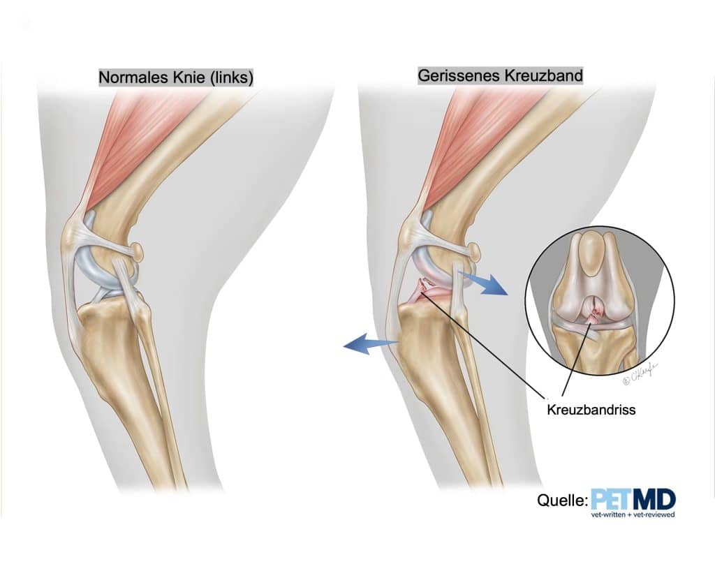
The basis of TPLO is that it prevents the instability called “shin thrust/drawer” that occurs when the anterior cruciate ligament in the knee joint is torn.
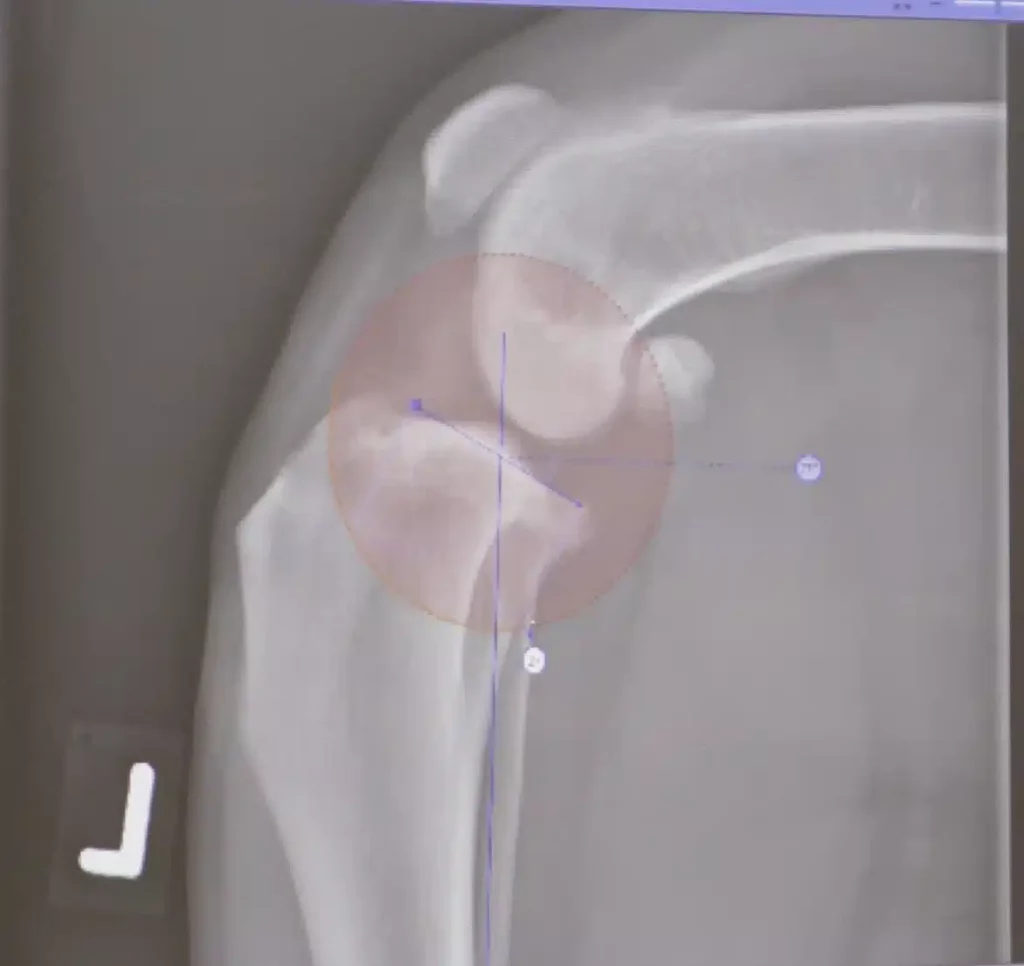
Unlike us humans, dogs have a sloping joint surface. This inclination of the tibial surface is usually between 20° and 30°, but in extreme cases it can even be greater than 40°. Like a cart on a sloping surface, the joint roller of the femur tends to slide down this slope. However, a healthy anterior cruciate ligament prevents this.

The anterior cruciate ligament is under a certain amount of tension with every step the dog takes. If the cruciate ligament is injured while jumping or running and begins to tear, it can no longer heal itself due to the constant tension. A cruciate ligament injury causes inflammation in the knee joint. Inflammatory enzymes (metalloproteases), which are produced in large quantities during joint inflammation, attack the already damaged cruciate ligament and articular cartilage. The cruciate ligament is then further weakened and osteoarthritis develops. Like a ship's rope rotting under tension, the anterior cruciate ligament tears completely at some point, usually when the knee joint is subjected to normal stress (“minor trauma”). At this point, advanced osteoarthritis in the knee joint can already be observed.
Please take a look at these chapters for the symptoms of a cruciate ligament tear.
TPLO – surgical method
During the operation, a quarter-circle bony incision is made at the upper end of the tibia. The articular surface of the tibia bone is then rotated backwards according to a previously calculated amount. It is fixed in this new position with a bone plate and screws.
The rotation reduces the inclination of the articular surface. The goal is a postoperative tilt of 5°. The angle between the patellar ligament and the articular surface is then approximately 90°. In experiments it was found that with this inclination the advancement of the shinbone is neutralized and the tension on the anterior cruciate ligament (if it has not yet been completely torn) is significantly reduced. Later arthroscopies performed two years after TPLO showed that in 16 of 17 dogs the torn cruciate ligament still existed and that the articular cartilage, menisci and all other joint structures were completely normal and healthy.
In order to avoid massive osteoarthritis of the knee joint, the operation should not wait until the cruciate ligament has completely torn. Ideally, TPLO is carried out when a cruciate ligament is torn in order to prevent further joint damage (osteoarthritis, meniscus tear).
Process of the TPLO operation
The most important thing in a successful TPLO operation is the veterinarian's experience as a surgeon with this surgical method and very precise planning. In addition, careful surgical hygiene is an essential prerequisite for an uncomplicated wound healing process.
On the day of the operation, the dog should be fasting, i.e. 12 hours without eating, and have successfully completed its walk while relieving itself. After a short general examination with particular attention to the ability of the dog to undergo anesthesia, a venous catheter is placed in the dog. After premedication with diazepam and painkillers, anesthesia is induced with a short-acting anesthetic (propofol).
The sleeping dog is then taken to the operating room preparation area where, after intubation, it is connected to inhalation anesthesia, anesthesia monitoring and intravenous infusion. The affected limb is now x-rayed in different planes. This is necessary for planning the operation.
After the X-ray examination, the patient is carefully shaved and washed. Depending on the size of the patient, approximately 2 hours have passed before the dog is transported to the operating room. In the operating room, the dog is tied up on the operating table and connected to the anesthesia system with artificial ventilation. A special intravenous antibiotic is now administered. The aseptically prepared surgical team drapes the dog with sterile surgical towels, the skin of the affected extremity is covered with sterile foil and the instrument table is prepared with all the necessary instruments.
Now the operation begins:
First, the knee joint is inspected. Particular attention is paid to meniscus damage, articular cartilage damage or other soft tissue injuries. The joint is then closed. The skin is then cut on the inside just below the knee joint and the TPLO is performed.
The dog is now taken from the operating room to the X-ray room or CT and follow-up X-rays are taken to measure the postoperative angle of the tibial plateau. Finally, the animal patient is taken to the inpatient department, where we look after him during the recovery phase. He is usually given a strong painkiller for the next 12 hours and the surgical wound is cooled with cold compresses.
The dog can be picked up by the owner in the evening - of course we will explain the process and the next steps and medication to you in a detailed final meeting.
Duration of healing process TPLO
In our experience, patients recover with better long-term outcomes than traditional passive stabilization techniques such as extracapsular lateral suture overlap. Just one month after surgery, patients can bear their weight well and are making progress with home physical therapy.
Possible complications of TPLO
Implant complications such as infection or screw loosening occur in a small percentage of cases and are treated with appropriate antibiotics and removal of the implant after healing.
Serious complications include: postoperative patellar luxation, tibial fracture, implant loosening/failure, and implant-related infections and meniscal tears (if the original meniscectomy/meniscus release was not performed).
Minor complications include : infection/inflammation of the incision (interface), seroma and wound dehiscence (pull apart of the edges of the wound).
TPLO operation in video
credentials
1 B Slocum, TD Slocum. Tibial plateau leveling osteotomy for repair of cranial cruciate ligament rupture in the canine . Vet Clin North Am Small Anim Pract (1993) 23:777–795.
